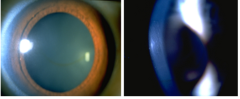
Confrontation and pupil testing is normal. Dilated fundus exam is also normal. Slit -lamp exam suggests some significant variation in corneal thickness; an optic section seems to be thinner inferiorly in the right eye and less pronounced in the left eye. Also you notice some vertical striae in the posterior cornea OS and a "greenish-brown" circular ring (see photos). You order corneal topography analysis.
Guiding Questions
1. What's an optic section?
2. Describe the anatomy and histology of the cornea.
3. What is the function of each layer of the cornea?
4. Why is the cornea transparent?
5. How does the composition of the sclera differ from the cornea? If the sclera is very thin (Stapholomata)
or a piece of it is dried out, it becomes transparent. Explain.
6. What controls the hydration of the cornea? What is the thickness-hydration relationship? Do the thin
areas seen by slit-lamp examination represent a change in hydration?
7. What are corneal striae?
8. What is the colored ring?